Medical Equipment Blog


Osteoporosis Model Q & A

Tuesday, May 31, 2011

I need a very small, highly compact osteoporosis model to take with me on rounds. What options are available?

A 4 stage osteoporosis model that is designed to be just four inches in diameter is the perfect option for easy portability. This model, is made of a series of discs that swing out to show a healthy, stage two, three and four cross section of bone and is just what is needed for easy transport. The benefit of these models is that they don't require one specific joint be affected, rather they highlight the changes in the bone itself. For use with a wide range of different patients and different health issues this is really a highly compact, very simple yet detailed tool for a medical professional to use.

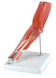
In working with patients with bone loss problems I have tried videos and charts and would like to try an osteoporosis model. Which model is the most comprehensive?

Depending on the specific location of the osteoporosis, there are several different models that highlight the progressive degeneration of the bone that occurs with the condition. The very small and highly compact disc model is perfect for any condition, but it is not three dimensional in nature. For an anatomical model that is in three dimensions you may want to consider a four stage vertebrae model or an osteoarthritis four stage knee model. This is a very comprehensive model that shows an exact yet slightly smaller version of the particular joint from every angle. The specific damaged done by osteoporosis, arthritis and other degenerative diseases is clearly visible by looking at the model. Combining a model with other types of information for the patient is highly effective. Continue using videos, pictures and written information along with the model. Each patient will use the information in different forms and will remember different presentations. Having a wide range of options can really help increase understanding.

Is there any osteoporosis model that shows the specific damage to the structure of the bone caused by the disease?

All high quality osteoporosis model options will show the specific damage to the bone, joint and surrounding tissues caused by the condition. Some models are very specific and highlight other features that can occur with the disease. For example the model of the spine will include marked bone compression as well as bone spurs. However having the other models also showing other spinal conditions can help in generating conversation and clearing up any misunderstanding about the diagnosis or the treatment.

Why is an osteoporosis model helpful in talking to an elderly patient?

Elderly patients may have difficult in hearing, comprehending and retaining information given to them about a specific type of diagnosis or treatment. Using an osteoporosis model to help the patient see what you are talking about is essential in ensuring that the patient is informed and knowledgeable about the condition. Not only can anatomy models be very effective with the elderly patient, but the spouse, caregiver, and family can also benefit from seeing the model and having a better understanding of the health condition.

Labels: , , ,


Veterinary Canine Models Q & A

Thursday, May 26, 2011

What options are available in skeletal types of veterinary canine models?
Veterinary canine models come in a wide range of options, and are most definitely not limited to just skeletal representations. There are many models that feature joints and skeletal parts, since these are commonly injured or diseased areas in dogs. The common joint models include the pelvis, elbow, shoulder and knee along with the vertebrae and the jaw. The jaw model is very realistic and is not the actual jaw bone itself, but rather the teeth in the gums in a realistic presentation. A clear canine jaw model is another popular option to show the adult canine teeth and their position in the mouth.

Other options for canine models that provide a lifelike view into a particular organ include the heart, heartworm model and the canine skin model. All are designed to be very accurate in their detailed presentation of the structures as well as the specific pathologies shown. In a vet's practice a two-sided canine ear model that shows inflammation and blockage on one side and a healthy dog's ear on the other is perfect for assisting owners in understanding the importance of routine hygiene programs for their pets.

I would like to use veterinary canine models in my practice to show my dog owners common disease conditions. Which ones are most popular and helpful?

Any and all of the veterinary canine models can be extremely helpful when working with pet owners. The skeletal anatomy models are perfect for discussions about the often inherited conditions such as luxating patella and elbow, hip dysplasia and arthritis in dogs. In addition the models of the canine shoulder, pelvis and vertebrae can also be used to show various conditions including trauma caused by falls, vehicle accidents or other types of injury. The information cards provided with the models give labels to each of the bones and structures allowing owners to follow along with the discussion. In addition the highly detailed 3D models also have the same type of educational card that may be used to provide more information about the specific healthy organ or the pathology displayed on the model.

One of the veterinary canine models that has a huge impact on owners is the heartworm model. This 3D model clearly shows the worms in the heart chamber exactly as they can occur in real life. This visual helps to keep owners using regular heartworm treatment in areas where this is a concern.

Are there the veterinary canine models that show pathologies or only those that show healthy joints and organs?
Depending on the manufacturer of the model, both healthy models as well as veterinary canine models depicting pathologies are available. Having both on hand to use with dog owners in general discussions about preventative treatments as well as in discussing treatment options and short and long term recovery is very helpful. Using the models help the owners to understand even very technical issues without a lot of explanation of anatomy and psysiology on your part.

Labels: , , ,


Colon Model Q & A

Wednesday, May 25, 2011

What pathologies are commonly depicted on a high quality colon model?

A high quality colon model will depict many of the common colon pathologies found in patient populations. These common health conditions include spastic colon, ulcerative colitis, diverticulosis, diverticulitis and Crohn's disease. In addition the best quality models will also show very simple colon pathologies such as bacterial infection, which can lead to many of the more significant issues if not resolved. Other diseases and conditions found on the model may include appendicitis and cancer.

Having a wide range of options on the anatomical model makes it very practical in using with any patient and his or her family. With more than one condition displayed on the model as well as sections of healthy colon comparisons are simple and easy to make. This type of model is also ideal for discussing long term management of chronic conditions such as Crohn's disease.

What models are a good compliment to a colon model in helping patients with a full understanding of digestive problems?
A full model of the entire gastrointestinal tract is an excellent compliment to the colon model. The larger model can be used to help patients to visualize the location in the abdominal cavity of the colon as well as its role in the overall digestive process. The smaller and more detailed colon model can then be used to get into the specific issues that the patient is experiencing.

For some conditions, a rectum model can also be used to help patients to understand the specific health conditions of the colon that are occurring in the internal or external rectal area. These can include internal and external hemorrhoids, annular cancer, abscesses, skin tags, fissures and cryptitis among other common and less commonly diagnosed health issues. Choosing to use either model in conjunction with the colon model will only help the patient to have a clearer picture of the condition as well as treatment options possible.

Is there are convenient to carry colon model option that can easily go from location to location or room to room as needed?
A very convenient to carry model that is easy to transport in a lab coat or examination kit is a four piece model that is attached to a backing by a hinge joint mechanism. Each part of the model can swing out to expose the internal features noted with that particular pathology. The pathologies depicted in the model include diverticulitis, chronic colitis, cancer and a normal colon section. The normal colon cross-section can be used as a reference and comparison section but also to talk about additional pathologies not depicted specifically in the model. The nice thing about these small sized models is that they also come with an educational card that provides specific information about each of the sections.

It is important to keep in mind that even the larger models are still very easy to move from room to room. The durable quality of these models is outstanding, and makes them a good match for any patient facility or teaching environment.

Labels: , ,


Thyroid Model Q & A

Tuesday, May 24, 2011

What are the common diseases shown on a thyroid model?

A good representation of the common thyroid conditions of Grave's disease, papillary carcinoma and lymphocytic thyroiditis, also known as Hashimoto's Thyroiditis, are the most commonly depicted medical conditions seen on a thyroid model. Having a normal, healthy thyroid model is also an important consideration for patient discussions and comparisons pre and post diagnosis. Education cards with information on the specific diseases depicted in the model are a great reference and also a good way for patients to read the main points and symptoms of the condition.

How accurately does a thyroid model show the differences between a healthy thyroid and one that is malfunctioning due to disease?
All thyroid models will not show the same level of detail when depicting either the healthy thyroid or a diseased thyroid. The best quality models will have clear and accurate depictions of both with no confusion as to the specific condition that is represented. Other features to look for in a very high quality anatomical model include the option to interchange the thyroid on the larynx to show patients exactly what the disease looks like internally. Having a card behind the model that shows the location of the larynx and thyroid in relation to the throat, chin and head is also very helpful for a patient to see the anatomical relevance of the model. Information on the card about each specific disease or condition that also highlights the differences will make the model a very effective teach tool in the examination room.
As the physician you can choose to share more information about the symptoms of the specific thyroid condition that the patient is experiencing. Using a variety of other models of the body can be helpful in drawing the line between the functioning of the thyroid and the condition or symptom being experienced by the patient.

Can a throid model be used with other models to explain to patients why they are experiencing different symptoms because of their thyroid condition?

Depending on the specific thyroid disease or condition there can be a wide range of associated health problems. Different joint and organ models can be used together with a thyroid model to allow patients to understand how a small gland in their neck is affecting other parts of their body. With all the various possible complications and symptoms seen in thyroid conditions other models that are good to use can include a wide variety of body parts. A hand model that show carpal tunnel and tendonitis is a good option for many patients. A female reproductive model is also useful since menstrual irregularities are associated with many thyroid conditions. Artery models that show plaque and cholesterol building up in the arteries and the possibility of partial or full occlusion of arteries due to these deposits are also highly recommended. For patients that are experiencing a wide range of symptoms an entire body model or an anatomical chart that links back to the thyroid model can be a great resource in explaining the variety of symptoms they are experiencing.

Labels: , ,


3D Anatomical Foot Model Q & A

Friday, May 20, 2011

I have been using an anatomical foot chart to help patients understand explanations, but I'm still not sure that I'm being really clear to some patients; is there another option?

Anatomical charts can be very effective in helping patients to visualize what is going on in their body as a system. For particular joints, organs and body parts the level of detail on a chart may not be enough. For a podiatrist or general practitioner that sees as a wide range of patients a 3D anatomical foot model can be a terrific tool to use when working with patients. Instead of using the chart as the primary source of visual information you can actually use the3D anatomical foot model as the primary aid in the conversation and use the chart to show how this condition may affect or impact other areas of the body. This is a natural progression of discussion from very specific information on the foot through to more general information on the rest of the body.

In a small office and practice what is the best options for a 3D anatomical foot model that doesn't take up much space?

A 3D anatomical foot model that is full size and mounted on a stand is not a large piece of equipment in any office or examination room. These models measure just 9 inches in length by just less than three inches in width and four inches in height. Surprisingly small, even with a small amount of the tibia and fibula, these models can easily sit on a desk or a counter. Many smaller offices keep the models in a central display area for patients to view. Other offices may keep the models in examination rooms for the same reason. Having these small mounted types of models is much less space consuming than a full human skeleton that is going to take up considerable floor and wall space.

What is the best way to wash a 3D anatomical foot model if it becomes dusty or dirty?

With routine use and exposure to a variety of different environmental materials a 3D anatomical foot model may begin to look a bit dismal and dull. Cleaning the model is very simple and requires just a soft cloth and some warm, soapy water. Start by gently wiping the model to remove any loose debris and dust. Then simply use a damp soapy soft cloth to wipe down the model. Follow this with a wipe with fresh warm water and allow to air dry. You will not need to use any type of cleaning solution but, depending on its use, you may also want to disinfect using standard disinfectants used on other plastic based hospital equipment.

In most situations with routine dusting by the cleaning staff there will be no requirement to clean the anatomy model. Since it is made of plastic it will not discolor, chip, peel or fade over time, which only adds to the value of the design.

Labels: , , ,


3D Human Brain Model Q & A

Thursday, May 19, 2011

Are there options for having a 3D human brain model combined with a system model for conditions such as hypertension and diabetes?

A full hypertension model or diabetes model will include a representation of the human brain. This style of 3D human brain model is designed to highlight the effects of these two conditions on the brain. This highlights the very real increased risk of stroke. The brain in these models shows the internal structure with the stroke occurring within one of the lobes of the brain. Very detailed anatomical representation of the brain is not common in these models since the goal is an overall understanding of the disease or medical condition. A good option is to combine a basic brain model with the model that shows all conditions associated with the specific disease. This allows for more detail when necessary without confusing the information supplied on the disease model.

What is the best type of 3D human brain model for an advanced anatomy class or laboratory?

An ideal 3D human brain model for advanced types of study is a brain that is located in the skull but also fully removable. This unique combination of brain and skull model provides the student will a good understanding of how the brain is located in the skull and where brain sections correspond to the outside of the skull itself. The brain model that is removable allows from maximum understanding of the healthy brain as well as the various common pathologies. If the brain can be removed and divided, a greater number of pathologies can be clearly and effectively represented. In addition details provided on an educational card that corresponds to the brain model provides further information of the pathologies depicted. This is an ideal study tool and one that is both interesting and engaging for student's to use in basic or advanced level anatomy and physiology classes.

I need a basic 3D human brain model that clearly shows the parts of the brain. Is there a detailed option that doesn't include any pathologies?

A basic 3D human brain model shows the right side of a human brain with all the external and major internal features. This is sometimes referred to as a sensory motor brain. Each component or part of the brain is colored differently to contrast and provide clarity as to the various sections. This brain is a normal size adult brain that is free from any pathology. The major focus of this model is to allow students and patients to clearly distinguish the various parts of the brain. This includes the lobes as well as the internal structures. Details include the olfactory bulb, optic nerve, optic chiasm, mammillary body and the medulla oblongata which are all associate with sensory functions.

This type of 3D human brain model is very beneficial in all types of educational settings as well as in working with patients. For example, using this model with stroke survivors is helpful in explaining the deficits they are experiencing as well as the options for rehabilitation and recovery.

Labels: , , ,


3D Human Lung Model Q & A

Wednesday, May 18, 2011

What are the most important features to look for in a 3D human lung model for a hospital emergency room setting for patient consultation?
Hospital emergency rooms are busy places with little room for storage and display. Finding a good match in a 3D human lung model takes a bit of time but there are definitely options available. Look for models that come mounted on a standard sized base that can easily be stored in a medical supply cupboard, under a counter, or on a counter itself. The smaller the model base and background card the less space the model itself will take.

A lung model for an emergency room setting should clearly indicate both health functioning as well as conditions such as asthma, bronchitis as well as chronic obstructive pulmonary disease or COPD. Most models will also show signs of lung cancer although in emergency room settings this diagnosis may not be common. Having both external and internal features of the lungs is also very beneficial for doctors and patients alike.

The model needs to be easy to clean and disinfect and also lightweight for easy movement between examination rooms. The model should also be designed to be durable and long lasting so buying from a well known and reputable anatomical model manufacturer is recommended for the best quality product.

What smaller models can be used with a 3D human lung model?

One of the most common small models that are used in conjunction with a full 3D human lung model is a bronchus model. This model is hinged at the back to a central rod that allows the cross-sections of the bronchus to swing out for display. It shows the detail of an asthma attack or chronic bronchitis where the passage swells and fills to prevent air passage. This bronchus model can be used together with the 3D human lung model to show those that have just experienced an asthma attack what happened in the body. They can also be used together to demonstrate how emergency inhalers and routine asthma medications work to keep lung passages open even when asthma triggers are present. By understanding this process patients can more accurately use emergency inhalers and stick to routine medications to prevent serious attacks that require medical treatment.

On a typical 3D human lung model what are the most common pathologies listed on the cards and depicted within the model itself?

There can be a wide range of diseases and conditions shown on a 3D human lung model. The highest quality models will show the greatest number of pathologies without resulting in a confusing depiction on the model. Different pathologies can be shown on different cut-aways and sides of the lung model for clarity and ease of viewing. Typical conditions noted on a good lung model include Chronic Obstructive Pulmonary Disease, asthma and cancer. The model can also be used to show fluid on the lungs associated with respiratory infections and other diseases.

Labels: , , ,


Artery Model Q & A


What options are there for an artery model for a patient examination room?
One of the best options for an artery model that is to remain in a patient examination room is a full model that is centered on a base. This model comes with a detailed educational card that sits behind the model and has an exact graphic replica of the model. On the illustration the various components of the artery as well as the effects of cholesterol and plaque deposits are clearly identified using correct medical terminology. Using this card helps the patient in following the doctor's information as well as in learning the actual terms used for the structures and conditions of the artery. These models are designed to be oversized with lots of detail to give the patient a very clear picture of the results of cholesterol and plaque in the circulatory system.

Is the same artery model used in a patient setting also a good option for using in a medical classroom?

One of the many benefits of using models of any type in a medical classroom is that students become very comfortable with the use of models. Students become familiar with the instructor and faculty using an artery model for medical explanations, helping the student to continue on with the presentation style when they are working with patients. A student that has experience in learning about cholesterol, plaque and artery damage through the use of an model will then see the educational benefit of using it with their patients.

The more that doctors are comfortable in using a variety of methods to share information with patients the better equipped they are to work with virtually anyone. The same types of anatomy model styles and types used in medical classrooms are very suitable for use with patients. Doctors can choose to provide as much or as little information to the patient as needed to ensure complete understanding of the patient's medical condition.

Is there a small, compact type of artery model that is perfect for doctors to use and easy to carry around on rounds or when working in a variety of examination rooms in a large clinical setting?

While a large oversized artery model complete with a stand and educational card is perfect for a desk, counter or examination room it is not always the best option to be carried to multiple rooms in a day. While there is always the possibility of having a model in each room there is still another choice as well. A small cylindrical model measuring just 1 3/8 inches in diameter and about 3 ½ inches long is perfect for slipping into a lab coat pocket on rounds or while working in clinical settings. This model works on a hinge pin that allows one or all of the four cross section pieces of an artery to be swiveled out for viewing. The model shows a normal artery at one end and a severely blocked artery on the other. The two middle rings show various degrees of cholesterol, plaque and diminishing diameter to the inside of the artery.

Labels: , , ,


Larynx Models Q & A

Monday, May 16, 2011

How specific are larynx models in showing the various complications and problems that can occur within the vocal cords and surrounding tissue?

Larynx models are not specifically designed to show the various complications and problems that can occur within the vocal cords and the resulting problems in vocalization. Instead, the model is designed to show the structure of the larynx and its relationship to the thyroid. The model will show the vocal cords on the model as well as the thyroid membrane and cartilage, cricoid cartilage, trachea and the hyoid bone. Understanding how the larynx sits in relation to other features of the airway and how it functions to allow sound is essential in understanding the consequences of different types of pathologies or damage. A doctor or speech therapist can use the model to inform the patient of the structural components that relate the larynx, which will important to understand the long and short term treatment goals.

What other anatomical features are present on larynx models?

The larynx, or voice box is a complex system of muscles, tissue and cartilage that creates air volume, pitch and sound and allows humans and other mammals to be able to create sound. The larynx is actually formed of a variety of features that are all shown on larynx models. These include but are not limited to the cricoid cartilage, thyroid cartilage, thyroid membrane, and the top of the structure, the hyoid bone. It will also show the trachea that forms the lower end of the larynx. The anatomy model will be an exact replica of a human larynx and anatomically correct from all perspectives. Very fine, identified parts of the larynx model will be highly visible even if they are not uniquely identified on the model. With additional study and in the case of specific conditions these additional features can be labeled or identified for patients.

Another major component of larynx models is the thyroid. The thyroid, which sits over the larynx at the base, controls the body's metabolic system as well as controls the production of a variety of hormones including growth hormone.

What other types of educational materials can be used in conjunction with larynx models to help patients to understand short and long term rehabilitation and speech therapy goals?
Many non-cancerous conditions of the larynx can be very successfully treated with speech therapy instead of surgical procedures. These include strains, overuse issues, muscle control problems and even air volume problems. Larynx models used in conjunction with posters and video presentations on how the larynx works and how muscles can be taught to correctly function are excellent options for a speech therapist.

Even in some medical conditions such as acid reflux or polyp removal surgery the use of speech therapy is typically recommended after the surgical procedure. The more information sources that are used to help the patient understand the short and long term goals of the therapy the more successful it is likely to be. Anatomical models are a great way to ensure that the patient has a thorough understanding of what is going on with his or her health.

Labels: , ,


3D Human Eye Model Q & A

Friday, May 13, 2011

Can internal features be viewed in a 3D human eye model?

All 3D human eye model types are designed to allow students, patients and researchers to look into the human eye. The cross-sectional models or the full eye that opens to reveal inner structures are perfect for both external and internal structure identification. Using the anatomical model with students and patients is an ideal way to allow them to see just what internal damage has occurred in the eye and why specific symptoms or types of vision loss occurs. Patients and students will both find that having a model to exam during presentation of the information helps to clarify and enhance understanding in a very natural and organized fashion.

All the internal features should also be found on a corresponding information card that is kept with the model. This card can be used to provide the medical names for structures in the eye as well as to provide additional information on a variety of eye conditions.

I want to show students a variety of different conditions of the eye that include the lens and cornea. Are there models available that provide this information?

There are four separate 3D human eye model options that feature a removable lens and corneas. The first two models are the basic and full eye model. These models have a healthy or normally shaped lens and cornea and are great for any type of basic anatomy class or medical study class. The second two models include the cornea eye cross-section and the cataract eye.

The cornea eye cross-section features four interchangeable corneas and corresponding information card. The card provides further information the result of the cornea malformation and how it impacts on vision and eye function. The cataract eye model has five interchange corneas that will help students understand the vision restriction that commonly occurs with cataracts of different degrees and types. The information card with this 3D human eye model highlights the damage to the cataract and specific conditions that may occur in vision or ongoing eyesight degeneration and possible permanent loss.

What is the best basic 3D human eye model for a classroom or lab setting?

Individuals that have the responsibility of ordering supplies and equipment for educational institutions have to be concerned with staying in budget as well as in buying high quality equipment. Buying directly from an anatomy model manufacturer typically is the best option since you are assured a high quality product that is developed to be accurate, durable and practical as well.

The basic 3D human eye model is no different that any other model. There are two models that are great for general study and they include longitudinal section model as well as a full eye model. They will both provide the same detail an information however the full eye model can be split open to reveal the interior structure while the longitudinal model will already display those structures. Both model options come with a removable lens and cornea and both are designed to be highly accurate in the depiction of the internal and external features of the eye.

Hip Joint Model Q & A

Wednesday, May 11, 2011

I deal with elderly patients. Is there a hip joint model you would recommend?

A basic hip model and an arthritic hip model are both good choices for working with geriatric patients. The basic hip model clearly shows the top of the femur as well as the hip joint, greater and lesser trochanter, ischial spine, ischium and pubis bones as well as the ilium. This provides an extremely accurate and simple perspective on the hip, ideal for discussing fractures, bone chips, deep muscle bruises and other conditions associated with falls.
The arthritic hip joint model is not made to be life sized. It actually shows four separate stages of arthritis in the hip joint. Even though each of the four representations is smaller than the basic hip they are very clear in showing the advancement or progression of arthritis in the joint. Erosion, bone spurs, fractures and degenerative joint disease are all displayed on the series of models. This can be instrumental in talking to elderly patients and their families about the aging process and what treatment options are available.

What is the difference between a basic hip joint model and a muscled hip joint model?

Both the basic hip joint model and the muscled hip joint are the same physical size with regards to height and width. The actual model of the muscled hip has additional features that include the gluteus medius, gluteus minimus, iliacus, inferior and superior gemellus, obturator internus, piriformis and psoas muscles. In addition the articular capsule ligaments are also clearly shown on the model. This type of model is ideal for teaching, research, and also for patient consultation and discussion. Having both the basic model as well as the muscled model is highly recommended in any type of health care setting. The two models compliment each other and allow the medical professional to choose the degree of detail they require in the model when working the patient.

What is the typical size of a hip joint model?

The more accurate in size that a hip model is, the more effective it is in helping the patient or student to clearly visualize an actual human hip. The best option when choosing a model is to look for a full size normal hip, which is typically going to measure approximately five inches by four and three quarter inches in length and width. Depending on the specific type of mounting to the base the height will be about eight and one quarter inches to stay accurate in dimensions. Both the basic hip and the muscled hip will have the same dimensions for a model size and the both will include the top of the femur to show how the ball joint of the hip works. These smaller sized, compact models are good for emergency rooms, patient care rooms in orthopedic units and in the office of a family doctor and take up much less space than a complete skeletal model or a full leg model.

Labels: , , ,


3D Anatomy Models Q & A

Tuesday, May 10, 2011

 What options are available for self-study with 3D anatomy models?

One of the best uses of 3D anatomy models is student self-study in laboratories and classrooms. The best models come with a handy card that can be mounted behind the actual model on the base. This card includes a graphic representation of the model, plus written information. Typically, most information will be the names of the various features of the joint, organ or body part. However, some models, including those that show cancer, disease or other medical conditions may also provide information on symptoms and related features. Good examples of this are the cancer models that provide additional information on the various stages of the disease within a particular organ or body part.

Are there very detailed anatomy models that show various types of conditions and diseases?

There are a wide range of 3D anatomy models that are specially constructed to clearly and realistically illustrate how disease affects different body parts and organs. These models are valuable both in teaching settings as well as working directly with patients. Different models may show the stages of cancers in various organs, the effects of osteoarthritis in the knee, arthritis of the hip or vertebrae degeneration.

Disease models are an excellent and very non-threatening way for patients and students alike to learn about specific conditions. While they are lifelike enough to be easily recognizable they are not anxiety provoking for the patient or the student to exam and ask questions about.

What is a good option for using 3D anatomy models with patients in consultation sessions?

Many doctors and medical staff choose to use 3D anatomy models as a regular tool when talking to patients of any age. The models can be placed on a table, in a cabinet, or beside the patient, and the medical professional can use the model as a guide to the discussion. Comparing what is going on with the patient to what is seen in the model is a good way to highlight both interventions as well as short and long term treatment goals. For patients that are hesitant to start treatment the models that depict the long term effects of the disease or the stages of the disease can be helpful in working with aggressive treatment options. Ultimately anywhere that a patient may not be completely clear on what is happening or what treatment will accomplish will benefit from the use of an anatomy model.

Would 3D anatomy models be a good idea in a therapy room or rehabilitation program?

Joint models are a very popular choice in 3D anatomy models for therapeutic settings. These models can depict healthy or damaged joints, perfect to increase awareness in the patient with regards to range of motion and mobility issues. Since many people are not experts on anatomy this added visual is a nice way to provide information without seeming to assume that the patient isn't knowledgeable. Using the models regularly when talking to patients further helps deal with this sometimes touchy issue.

Labels: , ,


3D Wrist Model Q & A

Monday, May 09, 2011

I work with patients with carpal tunnel syndrome and have a hard time explaining the condition. Would a 3D wrist model be helpful?
Carpal Tunnel Syndrome is one of the most common medical issues of the wrist and hand. Many have heard of the condition, but really don't understand just what it is. Trying to verbally explain the normal structure of the inside of the human wrist is challenging, but also having to explain when things aren't working correctly can be incredibly difficult. The good news is that a 3D wrist model can take most if not all of the stress out of this discussion. A high quality model will have the interior of the wrist facing forward, clearly showing the bone structure of the hand and wrist as well as the nerve bundle that runs through the wrist. Some models even provide musculature of the thumb, allowing you to further discuss problematic issues with patients.

Having the model available helps to put the patient at ease as well. They can point to things on the model to get addition information without having to try to remember medical terminology. This will help with overall communication and establish a great rapport with your patients.

Is a 3D wrist model labeled and how does that impact on the model's lifelike appearance?

A good quality 3D wrist model will not have labeling on the model itself. The labels do take away from the realistic appearance of the model plus the labels can be very difficult to read when printed this way. A better option is to have a separate card behind the model that clearly shows the names of the specific features of the model. A drawing that duplicates the model in front of the card is also used to help with the identification.

How long can I expect a 3D wrist model to last in a teaching environment?

With proper care a 3D wrist model will last for years even with daily use in a teaching environment. Look for durable, high quality plastic models that are well designed and manufactured. Plastic models are resistant to fading, chipping and pealing plus they can be easily cleaned with soapy water and a soft cloth. Handling the models will not cause damage provided that appropriate care is used.

Are 3D wrist models accurately sized for a normal human hand?

Most hand and wrist models are life-size and accurate for the typical human hand. They are also usually representing the right hand. Keep in mind that the 3D wrist model often looks slightly larger simply because there is no flesh visible, just the actual bones, tendons and nerves. Some companies offer the option of left hand models while still others may offer the option of custom design of models. Custom designed models are a good option for working with children or patients that have specific conditions of the hand or wrist. Arthritis models of the hand are a good example of this level of specialization.

Labels: , ,


Human Anatomy Models Q & A

Friday, May 06, 2011

What is the typical size of human anatomy models?
The specific size of human anatomy models varies greatly based on the type of model. For example, a model of the human eye is going to be much smaller than a model of the spine simply because of the size of the body part. Most body organ models will have a base size of approximately 61/2 inches by 5 inches. This compact size makes them easy to store on a shelf, bookcase, desk or on a counter. Most organ models will be life-size in design and the model itself will not exceed the edges of the raised base.
Large joint or spine models can measure up to 19 inches in length, especially models of the human elbow that include the arm, elbow, wrist and fingers. Foot models also tend to be slightly larger than the base. These models are centered on the metal rod that holds the model to the base. This centering means they are perfectly balanced and very steady and durable. Elongated models such as the spine feature specific areas of the spine, such as the lumbar region, and are also very balanced and durable. Entire spine, leg, arm or body models in life-size will be much larger, heavier and more challenging to store.
What care is required in moving and storing human anatomy models?

Human anatomy models are made of plastic, and are extremely durable and strong. They are also very lightweight and easy to carry or move from place to place. Generally there are no special care requirements for high quality human anatomy models. They can be cleaned using a soapy cloth with warm water if needed. The base and model can also be dusted as needed.
Through the use of state of the art plastics the colors and textures on the models do not degrade or diminish over time and use. The colors will not fade with exposure to natural or artificial light.
Are the most popular types of anatomy models readily available in most educational settings?

Most science, anatomy and biology classes will have at least a few human anatomy models on hand. However, there are also veterinary models that are perfect for vet training, basic animal first aid classes and comparative anatomy projects or class activities. Having a good variety of different styles and options in anatomy models can really add reality to the abstract and difficult to visualize elements of a classroom. Students of any age will enjoy working with the models and will benefit in being able to actually see a replica of the body rather than just looking at a diagram or picture.
How many human anatomy models per classroom are recommended?

The number of 3D models per classroom varies with the specific class subject matter as well as the level of study. Elementary classrooms often have only one model for class use while middle school to college classes may have models for groups of students to work on. Having at least one model in the class is beneficial, the more models that are available the more students will use them and enhance their learning.

Labels: , , ,


Knee Joint Models Q & A

Thursday, May 05, 2011

Do knee joint models just show the bones, or are there other features that can be found on different model options?

As with all types of models there are options for different features in knee joint models. The basic knee will include the bones as well as the major tendons and ligaments that hold the knee joint and knee cap in place. These ligaments typically fasten to the knee joint exactly how they would in an actual knee. With this level of detail most patient consultations are much easier and clearer, especially with patients that may have limited understanding of human anatomy.

Deluxe Anatomical Knee ModelOther options in knee joint models include the muscled knee model. As the name implies this is a model that includes the major muscles that are responsible for the movement of the knee. These muscles are very clearly identified as such and are a realistic color and texture. The muscles are located exactly where they are in the actual human knee and are very helpful in addressing knee sprains, strains and fractures.

I work in the emergency room. How practical is it to have knee joint models available for patients to understand what I am saying about their injury?

The emergency unit is a very fast paced and rather hectic part of the hospital or clinic. However, taking the time to clearly explain injuries or trauma to a patient is still foremost in a physician's mind. Having the option to grab a lightweight, easy to move model when the situation calls for a detailed explanation can actually save time, energy and decrease frustration levels on everyone's part. These models are designed to be very durable, easy to maintain and clean and also very lightweight for easy mobility as needed.

Most knee models are going to be small, about three inches in width and depth and ten inches in height. They can easily be stored in a central supply closet or at the nurse's station. When needed they can simply be brought to the emergency treatment room and returned after the patient discussion has ended. Doctors, nurses and even emergency room social workers can access the models as needed.

What is the benefit of having a card behind knee joint models and other types of human anatomy models?

Not all people viewing knee joint models are medical professionals or anatomy experts. Having a simple, clear and easy to read card located behind the model will allow anyone to identify the various components of the knee or respective body part. These types of models are ideal in patient rooms, in classrooms and even in research facilities. Having the features of the model identified is one option for the cards, but they can also be used to provide additional information about a specific condition or feature of the model. A good example of this is a breast model that provides information on how to do a self breast exam, which is an important part of preventative medicine for women.

Labels: , , ,


Shoulder Joint Models Q & A

Wednesday, May 04, 2011

I am in charge of inventory and supply. What features should I look for in shoulder joint models for a teaching and training focused hospital?

In any type of hospital or health care facility where training and ongoing professional development is a goal, high quality shoulder joint models are a great idea. They should also be combined with all other types of human anatomy models to provide a wide range of learning tools.
Anatomical Sports Shoulder Model
The key to finding the most comprehensive types of human anatomy models is to look for a company that has a good reputation and years of experience. Companies that have supplied pharmaceutical companies, doctors, health care facilities, clinics and schools of medicine are a great place to start. In addition models that include all the necessary features without crowding the model with labels and extraneous components are the best option. This allows the models to be a good option for working with staff as well as quick, simple and clear patient consultations.

Look for shoulder joint models that are designed to be durable, practical and realistic. These three features will ensure that the model is used frequently and becomes a part of how the staff interacts with patients and even with each other.

Are there any shoulder joint models that provide clear identification of all the parts of the shoulder?

There are different shoulder joint models with more or less detail depending on the type of model you require. Both options, the muscled or basic models, do have identification cards that are mounted to the back of the stand. These cards measure approximately the same size as the model and are a duplicate of the model in a smaller graphic representation. Each part of the model is labeled on the card with simple lines clarifying the various structures. The benefit of this type of model is that both the model and the card can be used to interact with patients, talk to clients or to share information between medical staff. The level of identification needs to be comprehensive on the card to cover any possible patient or teaching discussion that may occur.

Can I use shoulder joint models in combination with other types of teaching aids in my advanced anatomy classroom?

Models, including shoulder joint models, have a definite advantage over many of the more traditional types of teaching methods. Even in advanced level classes most students learn from reading text books or studying photographs or anatomical charts. While these teaching aids are helpful, combining them with time to study and examine a life sized shoulder model can reinforce all other types of learning. For highly visual learners that like the hands on approach models are an excellent way to study a body part from all directions, really increasing full understanding.

Models can also be combined with new types of technology such as 3d human anatomy software. The model can provide the physical presence while the online simulation or model can be used to supplement or compliment the learning process.

Labels: , ,


Elbow Joint Model Q & A

Tuesday, May 03, 2011

Elbow Joint ModelWhat is the typical size for an elbow joint model?

The elbow joint model is one of the many models that come in a variety of sizes. If you elect to have the simple or basic elbow joint only on the model the length of the model itself is 8.5 inches. The base is the standard 61.5in x 5in x 1in, which is ideal for storing on a shelf, counter or desk.  The elbow joint that includes the forearm and the hand is much longer, measuring in at 19 inches in length. Both options are very streamlined and compact, easy to store and move for classroom use or in patient rooms.  The full arm elbow models has the arm slightly bent to provide stability to the model and also allow easy viewing from a variety of different angles.

Are there different options in elbow joint model styles that provide additional detail over just the basic joint?

The basic human elbow joint model is great for teaching and working with students and patients alike. Although it is just the bone structure and corresponding ligaments it is specific enough for clearly demonstrating movement, muscle or ligament problems or fractures of the joint to patients. It is also perfect for anatomy students to use to learn how the hinge joint works.

Beyond the basic elbow joint model, there is the muscled elbow model. This model includes the musculature at the top of the elbow, clearly defining how those muscles work. Models will include the ligaments, the bones of the lower arm, wrist and hand as well as the major nerves that run from the upper arm through to the wrist. This type of model is good for both work with patients as well as classroom study and helps to show the working of the entire arm.

Where could I use an elbow joint model in a college level biology or general science class?

The elbow joint model is a great addition to any classroom because it appears so simple but is actually a very complex interconnection of the upper and lower arms. With major nerves running down the arm and through the elbow clearly visible in the model that reasons why the elbow can be so painful at times is very evident. Biology, science or anatomy classes can use the model to supplement the lecture as well as viewing pictures in their textbook.

Models with labeled cards that correspond to the features on the elbow or arm are a great way for students at any level to self-test and review. The cars can be removed during the review, allowing students to attempt to identify the various components of the joint. Students can then self-check their work by flipping up the card. This saves instructor time and also gives students a more active way to study anatomy. The models can also be used during examinations where numbers or letters are taped to the specific muscles, tendons, nerves or bones with the students required to supply the correct name for the feature. Elbow models can be used along with other anatomical and disease models for a thorough study of human anatomy.

Labels: , ,


Disease Models Q & A

Monday, May 02, 2011

What specific conditions are shown in disease models?
Many different disease models are available today to show everything from degenerative bone loss through to abnormalities of the prostate. Common models used in most hospital and health care settings include those showing stomach cancer, kidney disease, lung disease, breast cancer, colorectal cancer and skin cancer. However, other organs that are prone to specific types of cancers and diseases such as the liver, pancreas, gallbladder and spleen, colon and the digestive system are also represented with models. A hypertension model provides information on all the organs that this condition effects, really making a dramatic presentation to patients where this is a concern.

How do patients typically react to being shown a disease model? If I do use the model what should I expect?

Most patients react extremely positively to the use of disease models during a diagnosis or consultation. The models are lifelike and realistic, but they aren't as graphic or shocking as looking at actual body parts in photographs and videos. Since the patient is not negatively affected by seeing the model they are better able to hear and comprehend the information the medical staff is providing. The use of models may also generate more questions from the patient during and after the doctor's discussion. This is a very positive sign that the patient understands the disease and is focusing in on the consultation.

Are disease models helpful in assisting patients in understanding why early intervention is important in many types of diseases?

Many of the disease models are designed to show the stages or degeneration of the body over the course of a disease. This can be very helpful in showing patients the progression of the medical condition and what they can expect.  They can be very important in explaining the stages of cancer, osteoporosis, digestive disorders, arthritis and reproductive health issues. Having disease models to share with patients that may not understand that immediate or aggressive treatment is to their benefit is essential. The physician can use the model to create a visual impression of how the disease will progress. The patient can both see and hear the information which will help them make an informed decision. In some cases the models can be used with family members that may be making the decision on behalf of a patient.

Is it possible to have a custom made disease model?

Not all medical model companies offer the option to customize or create new and unique models based on a particular client's needs. The good news is that some companies do allow custom models, which can include disease models. For highly specialized practices, advanced medical classes and working with specific types of patients customized models may be just the answer. Custom models are typically more expensive than a stock type of model since they are unique and must be created from a one of a kind mold. Working with the medical model company in drafting and designing the model will be important to make sure you get just what you want.

Labels: , , ,